Arteriovenous Malformation
Arteriovenous Malformation (AVM) is a vascular malformation (abnormal network of blood vessels) where arteries shunt directly into veins instead of going through a bed of capillaries. This disfigurement of blood vessels lead to more blood flowing through, placing larger strains on the walls of the blood vessels. The walls of the blood vessels affected by AVMs are very often much weaker than normal vessels, and may cause ruptures (hemorrhages) from which blood leaks out, possibly causing damage to the surrounding areas. When this happens in the brain, it is called “Subarachnoid Hemorrhage,” which is life threatening, and a type of stroke. Unfortunately, it is also the way majority of diagnoses are made. AVMs are considered a “rare disorder,” with prevalence of around 0.02% (18 in 100,000), with no clear causes. They can occur in practically any part of the body with blood vessels, including the brain, face, stomach, and extremities.
The walls of the blood vessels affected by Arteriovenous Malformation are very often much weaker than normal vessels, and may cause ruptures (hemorrhages) from which blood leaks out, possibly causing damage to the surrounding areas.
Arteriovenous Malformation is a “rare” disorder, with a prevalence of around 0.02%, or 18 in 100,000 people (American Association of Neurological Surgeons). There are currently no well-established risk factors for AVM, but generally, it is regarded as a “developmental” or “congenital” (present at birth) vascular malformation.
Typically, one is born with an AVM, and as he grows older, the size of the malformation increases, but it does not “spread” as cancer may (Toronto Brain Vascular Malformation Study Group). Most are not inherited, with the exception of Hereditary Hemorrhagic Telangiectasia (HHT). An exception to congenital AVM is Dural Arteriovenous Malformation, which is generally acquired following an injury, and occurs in the dura (covering of the brain).
4 out of every 100 people with AVM will have a bleed during any one year (Toronto Brain Vascular Malformation Study Group)
Types of Arteriovenous Malformations
There are 4 types of AVMs:
- Arteriovenous Malformation (high pressure)– abnormal network of blood vessels, where arteries go directly into veins instead of going through a bed of capillaries.
- Cavernoma (low pressure)– abnormal cluster of enlarged capillaries with no significant feeding blood vessels
- Venous Malformation (low pressure)– abnormal cluster of enlarged veins (like spokes of a wheel) with no feeding arteries; rarely bleed, so are usually left untreated
- Capillary Relangiectasia (very low pressure) – abnormal capillaries containing enlarged sections
Based on Information Provided by the Mayfield Clinic
Brain AVM Grading System
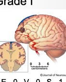 The AVM Grading System was developed in 1986 to estimate the risk of surgery. The scale anges from Grade I through V, based “on the eloquence of the surrounding brain tissue, venous drainage, and lesion size” (Barrow Neurological Institute). Please click on the image to find illustrated resource on the grading scale.
Peripheral AVM
A peripheral AVM is located outside of the head, neck and spine. It can occur anywhere, including the arms and legs, heart, lungs, liver and other abdominal organs, and even the reproductive or genital system. Read More